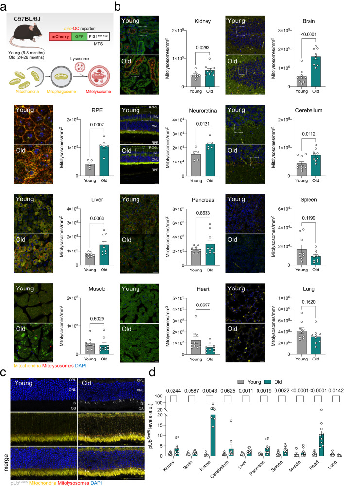
Fig. 1. Physiological aging in mice is associated with stable or increased mitophagy in multiple organs.
a Young (6–8 months) and old (24–26 months) mito-QC reporter mice bred on a C57BL/6J background were sacrificed and processed for confocal analysis. Created with BioRender.com. b Representative images and quantification of mitolysosome number (mCherry+GFP− puncta) in the kidney (renal cortex), brain (hippocampus), RPE, neuroretina, cerebellum, liver, pancreas (exocrine), spleen, muscle (gastrocnemius), heart, and lungs (n = 5–9 mice). Higher magnification insets are provided in Supplementary Fig. 1. c Representative images of whole eye cryosections from young and old mito-QC mice immunostained for phospho-UbiquitinSer65 (gray). d Quantification of phospho-UbiquitinSer65+ area in organs from young and old mito-QC mice (n = 5–9 mice). Scale bars, 25 μm (b) and 50 μm (c). All data are expressed as the mean ± s.e.m. Dots represent individual mice. P values were calculated using a two-tailed Student’s t test (b, Kidney, brain, RPE, neuroretina, cerebellum, liver, spleen, muscle, heart. d Brain, liver, spleen, muscle) or two-tailed Mann–Whitney U-test (b Pancreas. d Kidney, neuroretina, cerebellum, pancreas, heart, lung). Source data are provided as a Source Data file.

