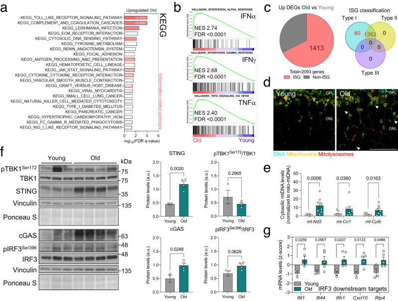
Fig. 2. Increased mtDNA release in old mice triggers cGAS/STING-mediated inflammation.
a Bulk RNA-seq data showing the top enriched KEGG pathways in old versus young retinas. Most correspond to inflammation-related signaling (color intensity corresponds to FDR q-value). b Top three enriched Hallmarks in old retinas: IFNα and IFNγ responses and TNFα signaling. c Interferome analysis (left) of upregulated DEGs in old retinas, showing the proportion of interferon-stimulated genes (ISG, 67.51%) and corresponding ISG classification (right). d Anti-DNA (cyan) immunostaining of whole eye cryosections from young and old mito-QC mice. Arrowheads indicate cytoplasmic DNA (GFP−DNA+) and the outer nuclear layer is shown. e qPCR measurement of retinal cytosolic mtDNA in young and old mice (mt-Nd2, mt-Co1, mt-Cytb), normalized to mtDNA content in the mitochondrial fraction (n = 8–9 mice). f Western blot analysis of cGAS/STING mediators (TBK1, IRF3, cGAS, STING) in young and old retinas (n = 3–4 mice). Protein levels are normalized to the loading control (Vinculin). g mRNA levels of downstream targets of the transcription factor IRF3 obtained from retina bulk RNA-seq data (n = 3–5 mice). Scale bar, 15 μm (d). All data are expressed as the mean ± s.e.m. Dots represent individual mice. P values were calculated using a two-tailed Student’s t test (e (mt-Nd2), f, g) or two-tailed Mann–Whitney U-test (e (mt-Co1, mt-Cytb). Source data are provided as a Source Data file.

