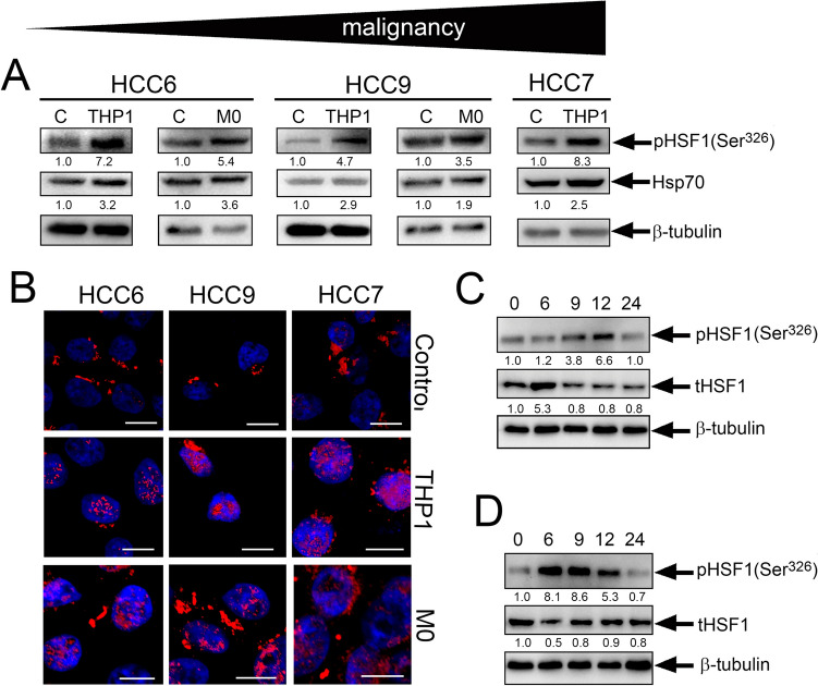
Fig. 4.
Pre-incubation of patient HCC cells with monocyte-like cells also leads to HSF1 activation. A Western blot analysis of HCC cells with anti-pHSF1Ser326 and anti-Hsp70 antibody after co-cultivation with THP1 or human monocytes for 20 h. β-tubulin was used as the loading control. The upper arrow demonstrates the level of malignancy of HCC cells according to medical diagnosis. B HCC6, HCC9 and HCC7 cells were seeded to cover glasses and then were incubated with THP1 or monocyte cells. Cells were stained with anti-pHSF1Ser326 antibody (red) and with DAPI (blue). Scale bar 5 µm. C The dynamics of HSF1 activation due to THP1 cells using the HCC6 cell as an example. HCC6 cells were co-cultured with THP1 at the indicated time intervals. D The dynamics of HSF1 expression after removal of THP1 from the co-culture at 20 h

