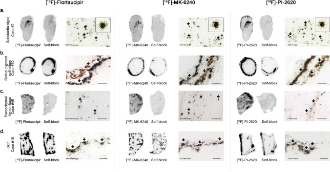
Fig. 4.
Representative images of [18F]-Flortaucipir (left), [18F]-MK-6240 (center), and [18F]-PI-2620 phosphor screen autoradiography experiments of slices containing substantia nigra in a control case (a), retinal pigment epithelium (b) in an dementia lacking distinctive histopathology case, parenchymal hemorrhagic lesions (c) and skin melanocytes (d) of an AD case. Strong binding of Flortaucipir, MK-6240 and PI-2620 was observed in neuromelanin-containing neurons of the substantia nigra (a), melanin containing granules in the retinal pigment epithelium (b), intraparenchymal hemorrhagic lesions (c) and skin melanocytes (d). Scale bars = 1 cm (a–d [18F]-Flortaucipir, [18F]-MK-6240 and [18F]-PI-2620 left panels: phosphor screen autoradiography) and 50 μm (a–d [18F]-Flortaucipir, [18F]-MK-6240 and [18F]-PI-2620 right panels; high resolution nuclear emulsion autoradiography)

