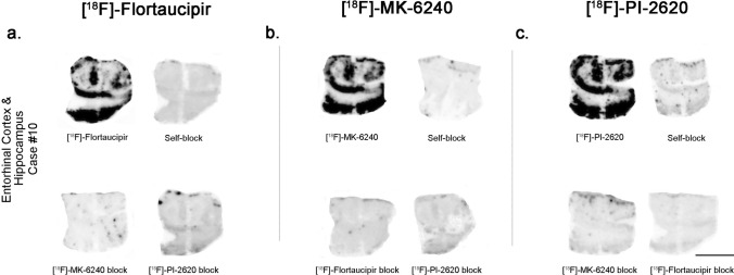
Fig. 5.
Head-to-head comparison of [18F]-Flortaucipir (left), [18F]-MK-6240 (center), and [18F]-PI-2620 phosphor screen autoradiographic binding patterns in adjacent sections obtained from the same tissue material containing entorhinal cortex from an AD case. The three tracers exhibited comparable strong binding to tangle-containing tissue material. Flortaucipir signal was almost completely blocked by adding 1 µM unlabeled MK-6240 or PI-2620; MK-6240 signal was almost completely blocked by adding 1 µM unlabeled Flortaucipir or PI-2620; and PI-2620 signal was almost completely blocked by adding 1 µM unlabeled Flortaucipir or MK-6240. Scale bar = 1 cm

