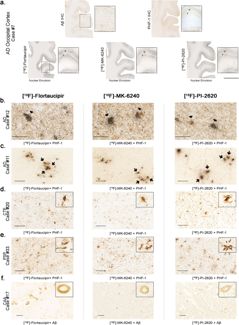
Fig. 7.
Representative [18F]-Flortaucipir (left), [18F]-MK-6240 (center), and [18F]-PI-2620 (right) high resolution autoradiography photomicrographs of brain slices containing occipital cortex from AD (a, b), temporal cortex from CTE (c), basal ganglia from PSP (d) and occipital cortex from CAA (e) and immunostaining with appropriate antibodies [PHF-1 antibody (kind gift of Dr. Peter Davies) and anti-Aβ antibody (1:500, mouse, clone 6F/3D, Dako)]. High resolution nuclear emulsion autoradiography showed a strong cortical accumulation of silver grains with the three tracers in cortical layers III and V (a) in AD brains mirroring the laminar pattern of tangles on adjacent slices as revealed by PHF-1 immunostaining rather than the more scattered plaque distribution pattern revealed by Aβ immunostaining (a). Strong silver grain accumulation was observed coinciding with the location of phosphor-tau immunoreactive cell somas (arrowheads) (b) and neuritic dystrophies (arrows) (c) in AD. No silver grains could be detected co-localizing with any of the three tracers in tau aggregates in CTE (d) or PSP (e) or with vascular amyloid deposits in CAA (f). Abbreviations AD, Alzheimer disease; CAA, amyloid angiopathy; CTE, chronic traumatic encephalopathy; PSP, progressive supranuclear palsy. IHC, immunohistochemistry. Scale bars = 1 cm (a), 50 μm (b), 100 μm (c–e), 200 μm (f)

