
Figure 5: Reduced recruitment of functional brain networks during evidence integration in schizophrenia patients.
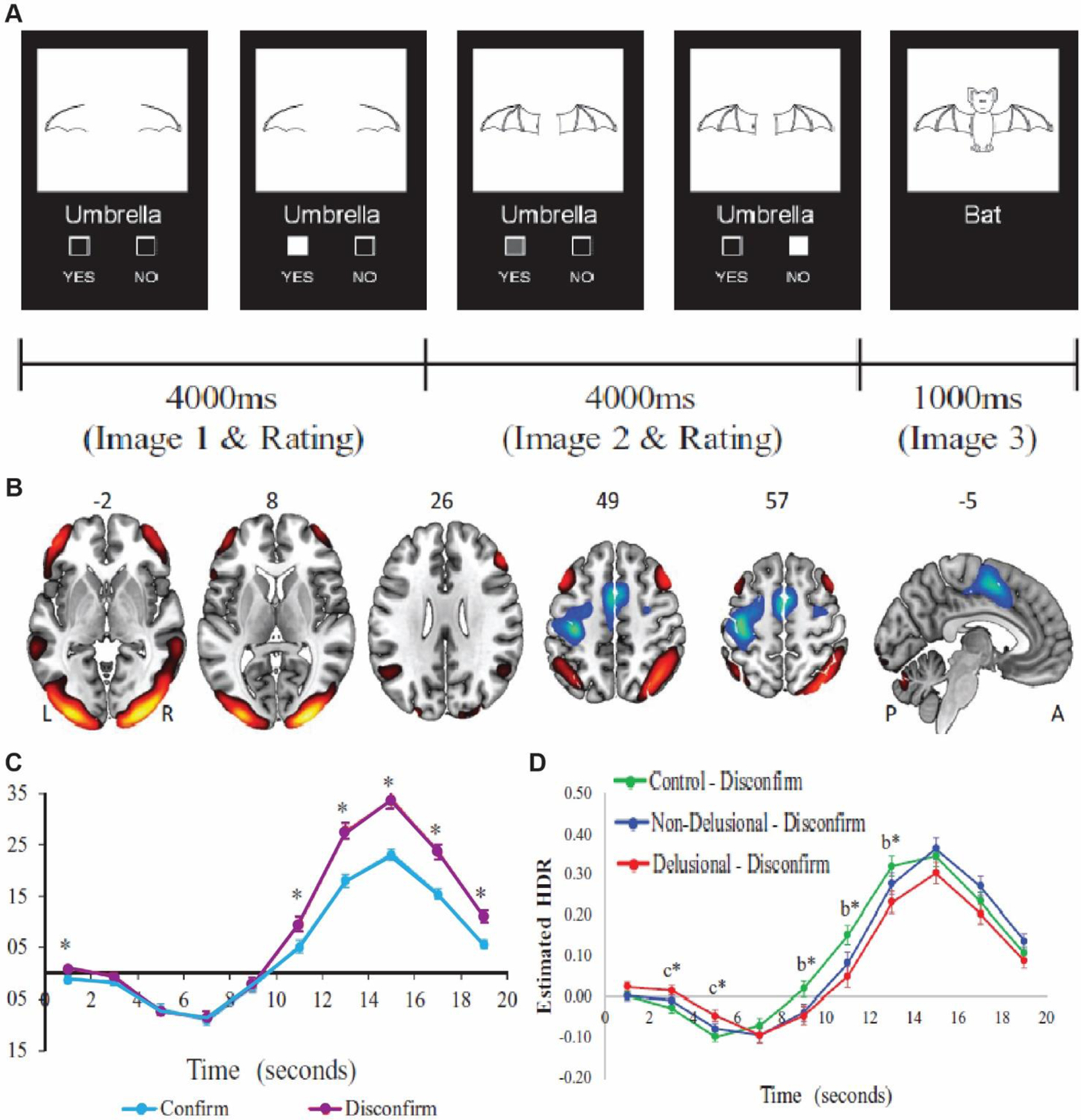
(A) In this task participants are presented with two consecutive partial line drawings of an object and a word describing it, with the second picture being more complete. Subjects were asked if the displayed word described the full picture upon presentation of the first picture and then to confirm/unconfirm, their decision when presented with the second picture. (B) Task induced activation of the cognitive evaluation network (CEN) comprised of bilateral orbitofrontal, dorsolateral PFC, angular gyrus, midline temporal gyrus and occipital lobe. (C) The CEN recruitment measured through fMRI is higher for disconfirmatory evidence integration (purple) than for confirmatory ones. (D) Schizophrenia patients with delusion (Red) show lower activation of the CEN network compared to controls (green) as well as non-delusional patients (blue). Adapted from Lavigne and others 2020.