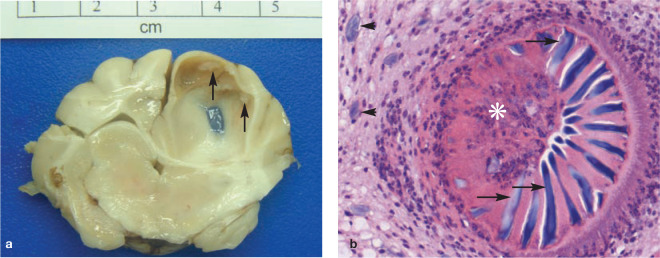
Figure 3.
Cerebral coenurosis in a Birman cat. (a) The inner surface of a large parasitic cyst within the left parietal lobe is multifocally roughened (arrows) corresponding to aggregations of invaginated protoscolices arranged in an approximately linear fashion. (b) Protoscolex (asterisk) showing the rostellum armed with chitinised hooks (arrows). Note the basophilic calcareous corpuscles (arrowheads) in the adjacent parenchyma. H&E stain, x 400 magnification

