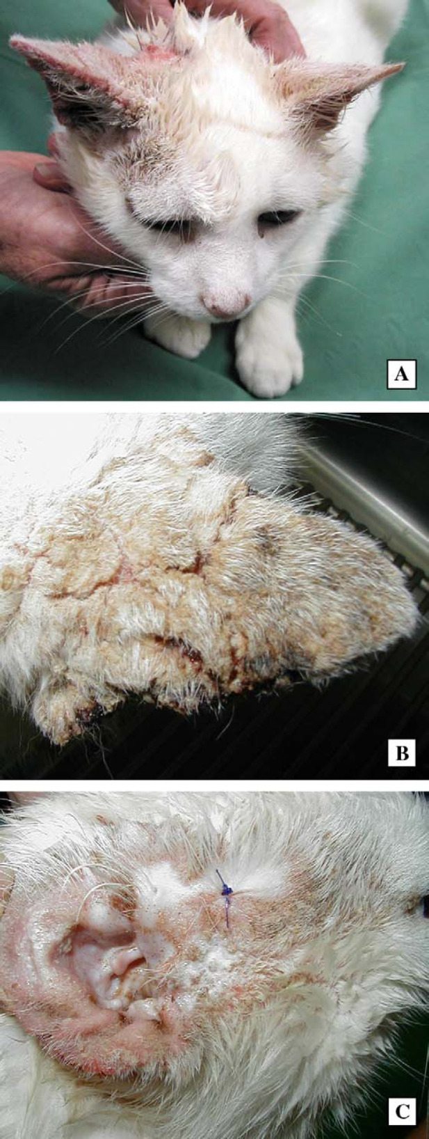
Fig 1.

Photographs demonstrating the lesions present in case 1. The most conspicuous lesion is the crusting fissured dorsal surface of the pinna (A), seen most clearly in the second photograph (B). Similar lesions, but of lesser severity, are evident on the ventral surface of the pinna and on the periorbital area of alopecia (C).
