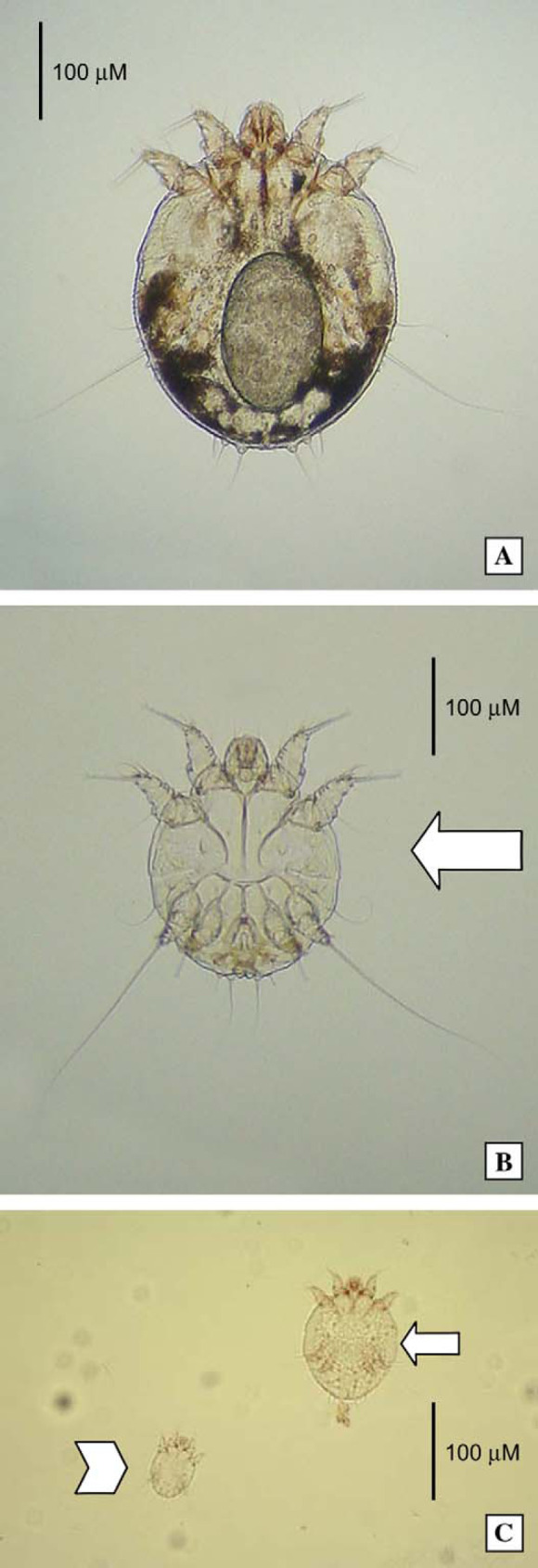
Fig 3.

Photomicrographs of sarcoptic mites from case 1, at higher magnification. A female mite, containing an egg, is present in the top micrograph (A). A male is identified by the large arrow in the middle photograph (B), while a nymph (small arrow) and hexapod larva (arrow head) is seen in the lower most image (C). (A) and (B) original magnification − 100×; (C) original magnification − 50×.
