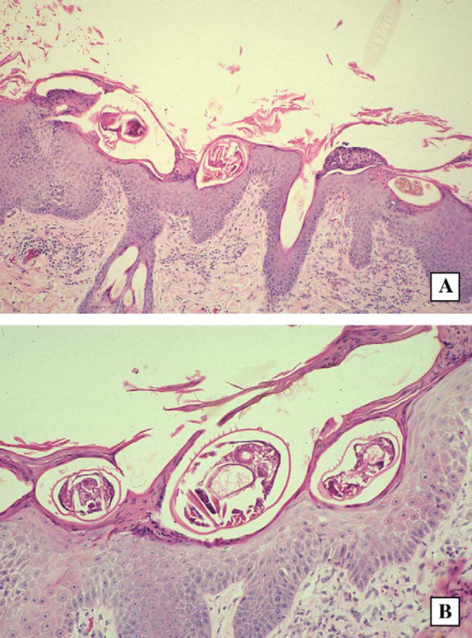
Fig 4.

Histological sections from case 1, a cat with crusted scabies. Note the parakeratosis, hyperkeratosis, presence of mites in epidermal ‘tunnels’ or burrows and mild inflammatory infiltrate in the dermis. H&E; original magnification − 40× and 80×, respectively.
