Fig 8.

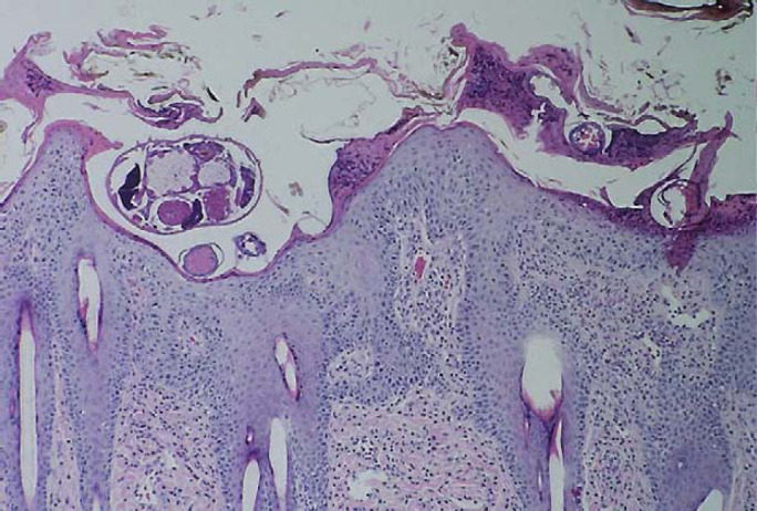
Photomicrograph of skin biopsy specimen from case 3 showing hyperplastic spongiotic crusting with numerous eosinophils. A cross section of a Sarcoptes scabiei mite is evident in the superficial epidermis. H&E; original magnification − 40×.

Photomicrograph of skin biopsy specimen from case 3 showing hyperplastic spongiotic crusting with numerous eosinophils. A cross section of a Sarcoptes scabiei mite is evident in the superficial epidermis. H&E; original magnification − 40×.