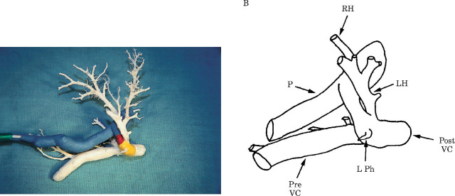
Fig 5.
(a) A painted corrosion cast of the hepatic vasculature from a cat with a left divisional shunt consistent with a PDV, photographed from the left lateral apsect. (b) The cast is also represented as a line diagram. The same colour codings and annotations apply as to Fig 4.

