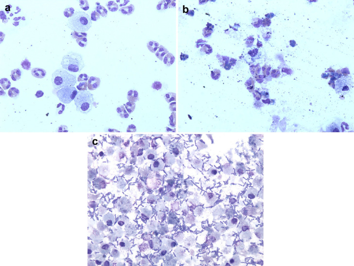
Fig 1.
(a) Photomicrograph of BALF processed within 1 h of collection (baseline) assigned a morphology score 1 and bacterial score 1. More than 90% of the cells are easily identifiable and there is no evidence of bacteria. Wright–Giemsa stain; original magnification ×600. (b). Photomicrograph of BALF stored at RT for 48 h assigned a morphology score 4. There is a large amount of amorphous debris and it is impossible to accurately identify more than 75% of cells. Wright–Giemsa stain; original magnification ×600. (c). Photomicrograph of BALF stored at RT for 48 h assigned a bacterial score of 4. There are abundant extracellular bacteria noted. Wright–Giemsa stain; original magnification ×600.

