
Figure 1.
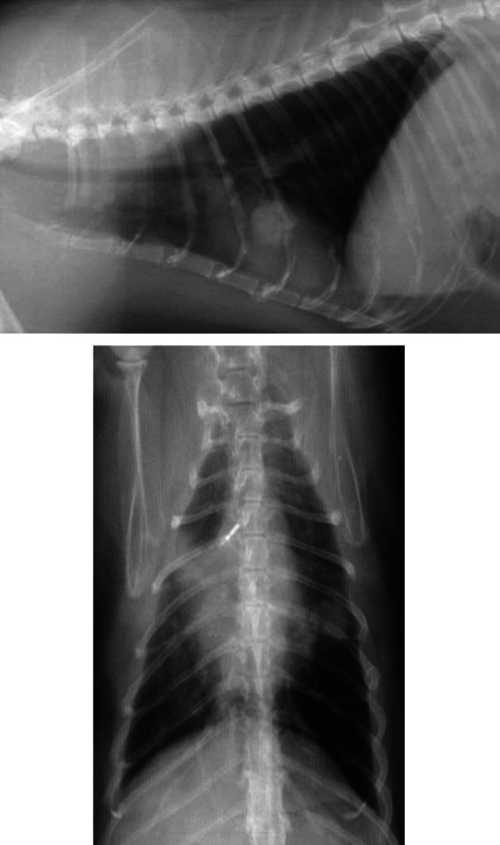
Right lateral and ventrodorsal radiographs from Case 3 (mycoplasmal abscess). A well circumscribed opacity is superimposed upon the cardiac silhouette at the level of the fifth and sixth intercostal space (Figure 1a). It has clearly defined margins and matches the soft tissue opacity visible in the caudal part of the left cranial lung lobe (Figure 1b). An ill-defined pulmonary opacity in the right cranial lobar region is also evident (Figure 1b).