
Figure 2.
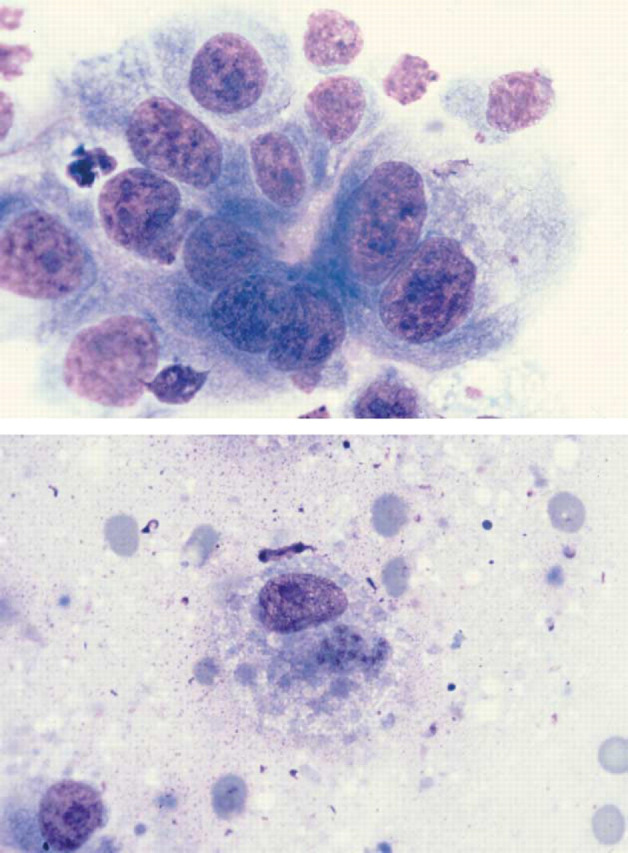
(a) Diff Quik-stained squash preparation of pulmonary parenchyma from Case 21 illustrating dysplastic bronchoalveolar epithelial cells. Note the variation in cell and nuclear size, the presence of prominent large nucleoli and one binucleate cell. Magnification ×735. (b) T. gondii tachyzoites within a macrophage in a squash preparation of pulmonary parenchyma from Case 21 (Diff Quik stain). Magnification ×735.