
Figure 3.
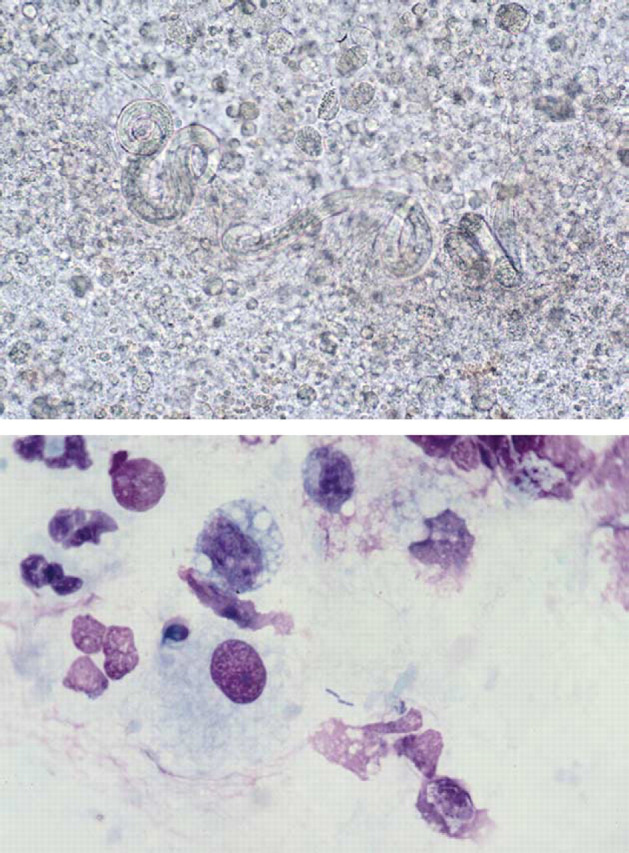
(a) Unstained wet preparation of the bronchoalveolar lavage from Case 17 illustrating larvae of A. abstrusus surrounded by inflammatory cells. Magnification ×147. (b) Diff Quik-stained smear of bronchoalveolar lavage from the same case illustrating bacterial rods of S. typhimurium surrounded by bronchoalveolar macrophages and intact and degenerate neutrophils. Magnification ×735.