
Fig 3.
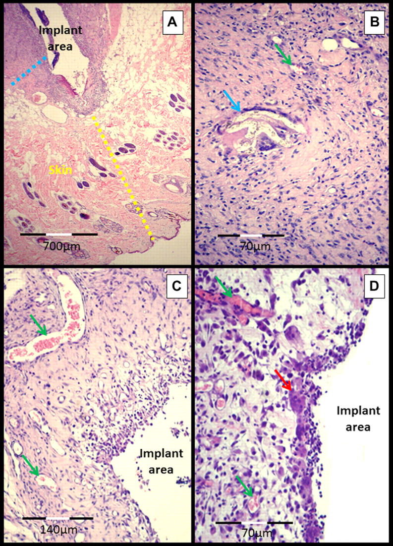
Micrograph of the subcutaneous–implant interface 45 days after experimental surgery for the implantation of a 70% PHB and a 30% HA composite in the subcutaneous tissue of cats. (A) Skin (yellow dotted line), subcutaneous tissue and fibrous capsule over the implant (blue dotted line). (B) Fibrous capsule in greater detail, showing the biomaterial (birefringent appearance) inside a giant cell (blue arrow), the green arrow points to a vessel. (C) Fibrous capsule with intense neovascularization (green arrows) and multinucleated giant cells (red arrow).