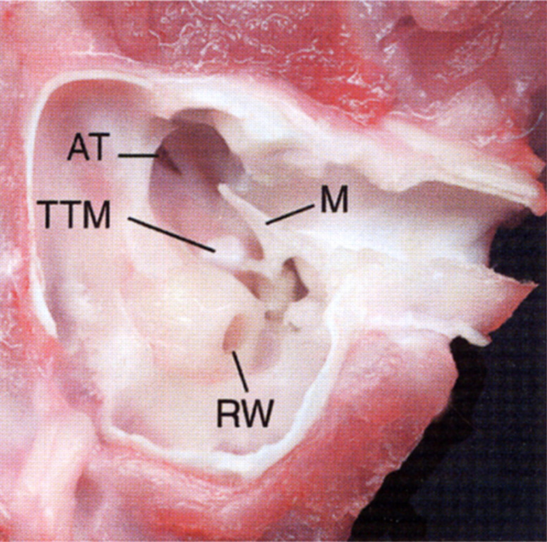
Fig 5.

Ventral view of an anatomical dissection of the left tympanic cavity of a cat. The tympanic bulla and bony septum have been removed to expose the epitympanic recess. Note the round window (RW), malleus (M), tensor tympani muscle (TTM), and opening of the auditory tube (AT).
