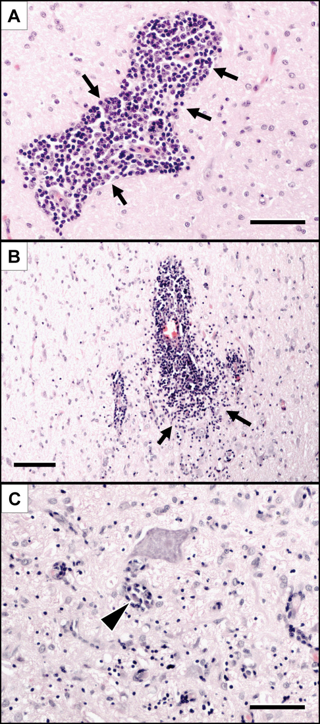
Figure 1.

Histopathological changes observed in the brain of the cats included in this study.
Lymphohistiocytic infiltrates (arrows) are either confined to the Virchow-Robin spaces (A) or diffusely invade the perivascular neuroparenchyma (B). Further immune cells are occasionally centred on degenerating neurons (C, arrowhead). Haematoxylin-eosin stain. Scale bars: A and C: 50 µm; B: 100 µm
