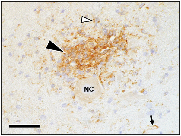
Figure 3.

The area of infiltration intensely stains immunopositive for Mx-protein (black arrowhead). The protein is expressed mainly in immune cells and astrocytes, including their cellular processes (white arrowhead). Moreover, the antigen is present in endothelial cells of intra- and perilesional brain capillaries (small arrow). Chromagen: diaminobenzidine (brown) with haematoxylin counterstain (pale blue). NC = nerve cell. Scale bar: 50 µm
