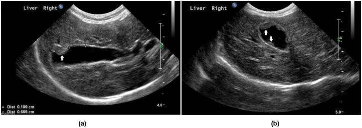
Figure 1.
Ultrasound images of the gall bladder of a cat presented with a mild decrease in appetite and increased liver enzymes. Longitudinal (A) and transverse (B) views are shown. Multiple sessile hyperechoic structures, indicated by white arrows, were noted along the luminal aspect of the gall bladder wall. The largest nodule measured 0.7 cm long (indicated by ‘x’ caliper marks on the longitudinal view). The thickness of the remainder of the gall bladder wall was unremarkable (indicated by ‘+’ caliper marks in the longitudinal view)

