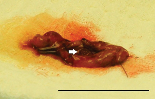
Figure 2.

Photograph of the excised gall bladder of a cat presented with a mild decrease in appetite and increased liver enzymes. Multiple nodules within the gall bladder wall were palpated during surgery and visualized upon dissection after cholecystectomy. The white arrow indicates one of the nodules visualized on the luminal aspect of the gall bladder wall. The black reference bar measures 4 cm in length
