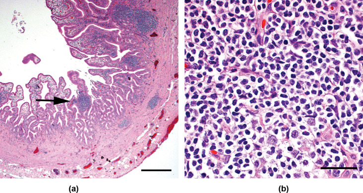
Figure 3.
Photomicrographs of the gall bladder wall of a cat presented with a mild decrease in appetite and increased liver enzymes. Multifocal areas of infiltration with small, well-differentiated lymphocytes were noted. The black arrow in (A) indicates one area of lymphocytic infiltration, which is shown at greater magnification in (B). The black reference bar in (A) measures 400 µm in length; the reference bar in (B) measures 30 µm in length. The sample was stained with hematoxylin and eosin (H&E) stain

