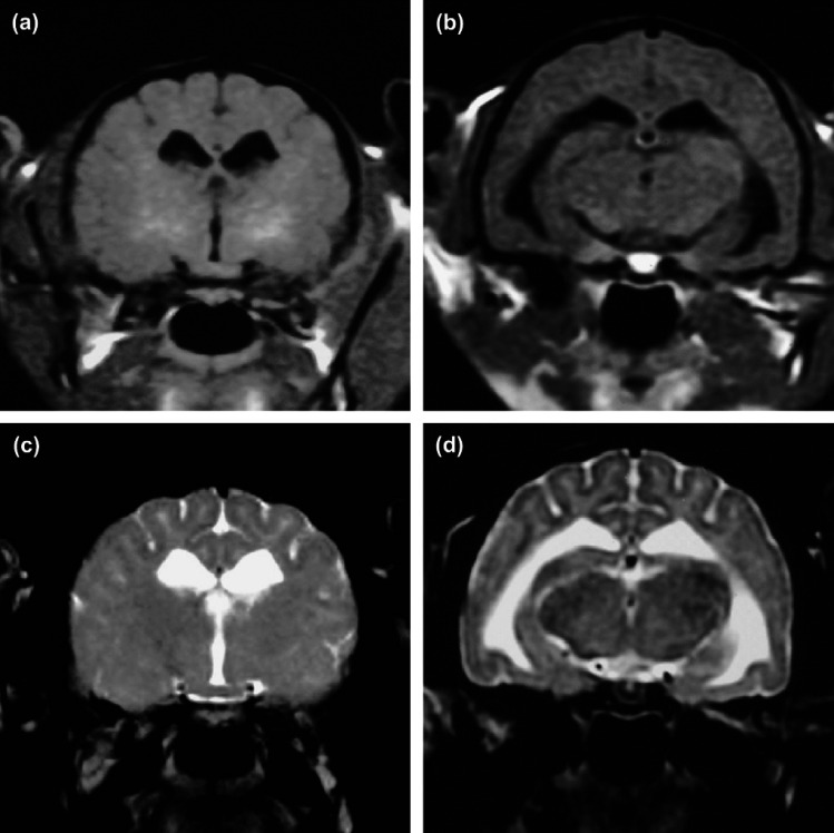
Fig 2.
Magnetic resonance imaging of a kitten (case 2) with GM2 gangliosidosis. (a, b) Transverse T1-weighted imaging at the level of the thalamus and hippocampus. (c, d) Transverse T2-weighted imaging at the same level as (a) and (b). Mild hyperintensity on T1-weighted image in the internal capsule (a) and ventricular enlargement (b), and hyperintensity on T2-weighted image in the white matter of whole forebrain (c and d).

