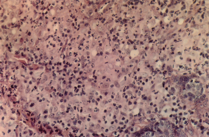
Fig 1a.
Haemotoxylin and Eosin (x100). Histological examination of 5 μm sections stained with haematoxylin and eosin (H&E) showed expansion of the dermis and subcutis by a mass of pyogranulomatous inflammation. The histiocytes have a moderate amount of pale staining to eosinophilic cytoplasm and have hypochromic nuclei, often with a single nucleolus.

