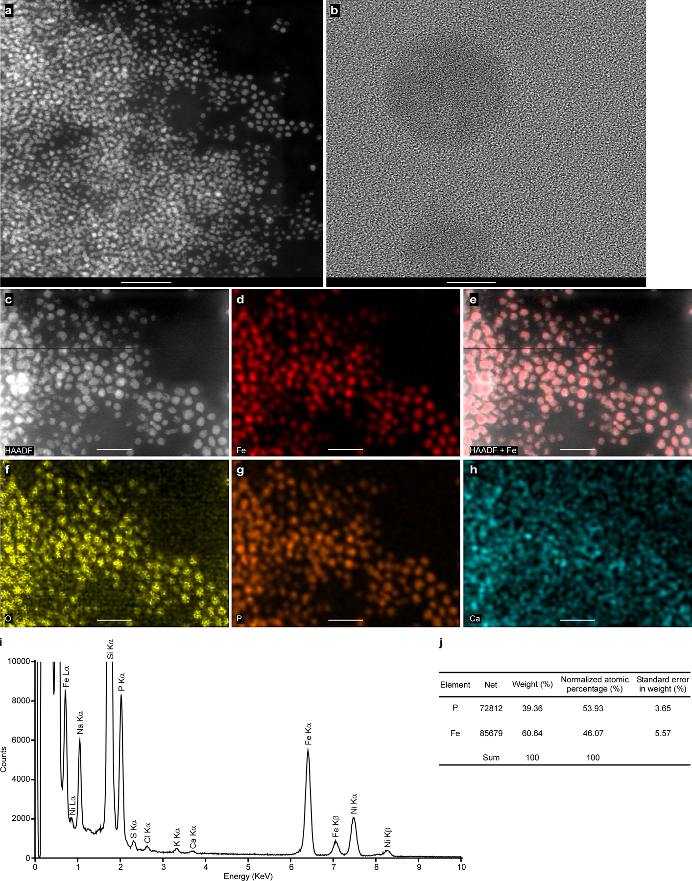
Extended Data Fig. 5 |. Isolated ferrosomes are amorphous iron- and phosphorus-rich granules.

Images shown are (a) HAADF-STEM, (b) TEM, no crystalline structures were observed in the isolated ferrosomes, (c) HAADF-STEM, (d) Fe EDS map, (e) HAADF merged with Fe signal to show colocalization of Fe signal with electron-dense granules, and EDS maps for (f) O, (g) P, and (h) Ca. (i) The integrated EDS spectrum of the isolated ferrosomes shown in panel c. Three distinct Fe peaks (Lα, Kα, and Kβ) were observed but no other transitional metals were detected above background levels. Some background signal of Ca was detected. The samples were deposited on 200 mesh Ni TEM grids and strong Ni peaks were detected. Scales bars, (a) 200 nm, (b) 10 nm, and (c-h) 100 nm. ( j) Elemental quantification based on the EDS spectrum of the isolated ferrosomes. The ratio of P versus Fe is 54:46. Experiments were conducted at least five times with similar results.
