
Extended Data Fig. 8 |. The entire fez operon is required for ferrosome formation in B. subtilis 168.
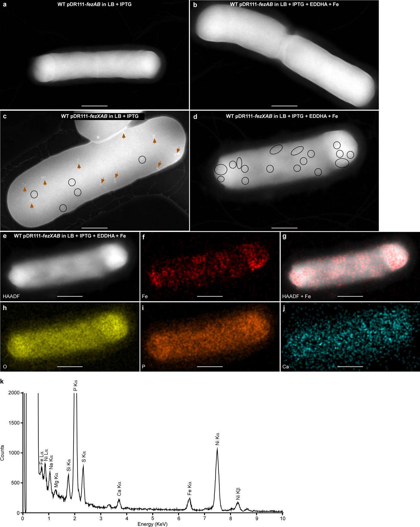
(a-b) Micrographs of B. subtilis cells expressing fezAB (WT pDR111-fezAB). No electron-dense ferrosomes were observed. (c-d) Micrographs of B. subtilis cells expressing the entire fez operon (WT pDR111-fezXAB). Electron-dense granules are circled, and disordered iron precipitates are denoted by orange arrows. (a and c) Cells were grown anaerobically in LB; (b and d) Cells were grown anaerobically in LB amended with 100 μM of the iron chelator EDDHA for 4 h, followed by addition of 500 μM FeSO4 and 3 h incubation. IPTG (1 mM) was used to induce expression of either fezAB or the fez operon. (e-j) Representative micrographs of the same cell in panel d by (e) HAADF-STEM or (f-j) EDS maps of (f) Fe, (g) HAADF merged with Fe signal, (h) O, (i) P, and ( j) Ca. Scales bars, (a-d) 500 nm and (e-j) 400 nm. (k) The integrated EDS spectrum of the micrograph in panel e. Two distinct Fe peaks (Lα and Kα) were detected but no other transitional metals were detected above background levels. The bacterial cells were deposited on 200 mesh Ni TEM grids, and strong Ni peaks were detected. Experiments were conducted at least three times with similar results.