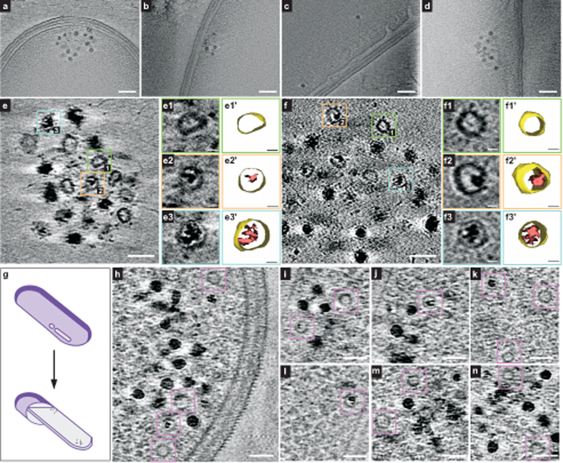
Fig. 4 |. Ferrosomes are membrane-bound iron storage organelles visualized by cryo-ET.

a–d, Four representative tomographic slices show ferrosome clusters localized in proximity to the cell membranes of intact fur::CT cells. The full video of the reconstructed electron tomograms is in Supplementary Video 2. e,f, Tomographic slices show that isolated ferrosomes are membrane bound. Boxed areas mark empty vesicles and vesicles filled with varied levels of iron phosphate minerals. Magnified views (e1–e3, f1–f3) and 3D segmentation models (e1′–e3′, f1′–f3′) of membrane vesicles are shown. The full videos of the reconstructed electron tomograms are in Supplementary Videos 6 and 7. g, A schematic of cryo-FIB milling. h–n, Seven representative cryo-electron tomographic slices in FIB-milled lamellae (around 200 nm thick) show that ferrosomes are membrane bound. Purple boxes mark empty vesicles and vesicles filled with varied levels of iron phosphate minerals. The full videos of the reconstructed electron tomograms are in Supplementary Videos 8–10. Scale bars, 100 nm (a–d), 50 nm (e,f), 10 nm (e1–e3, e1′–e3′, f1–f3, f1′–f3′), 50 nm (h–n). FIB, focused ion beam.
