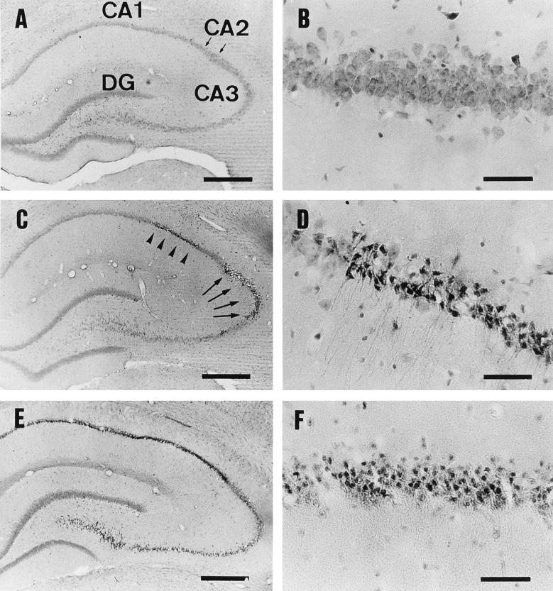
FIG. 2.
HE-stained sections of the hippocampus. Rats were injected with the vehicle alone (A and B), 50 ng of epsilon-toxin per kg (C, D, and F), or 100 ng of epsilon-toxin per kg (E) and then sacrificed at 4 h (A through E) or 24 h (F) p.i. (A) The areas denoted CA1, CA2 (an area between two small arrows), CA3, and DG are the CA1, CA2, and CA3 subfields and the dentate gyrus in the control injected with vehicle alone, respectively. (B) Higher magnification of panel A, showing the appearance of intact pyramidal cells in the CA1 subfield. (C) Appearance of the hippocampus at 4 h p.i. with 50 ng of epsilon-toxin per kg, showing the damage to the CA1 (arrowheads) and CA3 (arrows) subfields. (D) Higher magnification of panel C, showing so-called dark cells in the CA1 subfield. (E) Appearance of the hippocampus at 4 h p.i. with 100 ng of epsilon-toxin per kg. Note that almost all of the pyramidal cells, but not the dentate gyrus, are damaged. (F) Appearance of pyramidal cells in the CA1 subfield at 24 h p.i. with 50 ng of epsilon-toxin per kg. Note that the dark cells have transformed into eosinophilic cells. Bars, 500 μm (A, C, and E) and 50 μm (B, D, and F).

