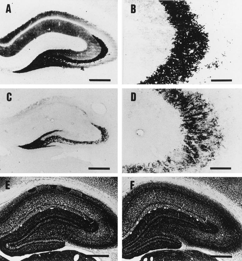
FIG. 4.
Timm’s zinc staining (A through D) and AChE staining (E and F) of the hippocampus. Rats were injected with the vehicle alone (A, B, and E) or 50 ng of epsilon-toxin per kg (C, D, and F) and then sacrificed at 4 h p.i. Panels B and D are higher magnifications of the CA3 subfields in panels A and C, respectively. Note that epsilon-toxin intoxication decreased the intensity of zinc staining in the hippocampus (C), especially in the mossy fiber layers of the CA3 subfield (D), while it did not change the density or distribution of AChE-positive fibers (E and F). Bars, 500 μm (A, C, E, and F) and 100 μm (B and D).

