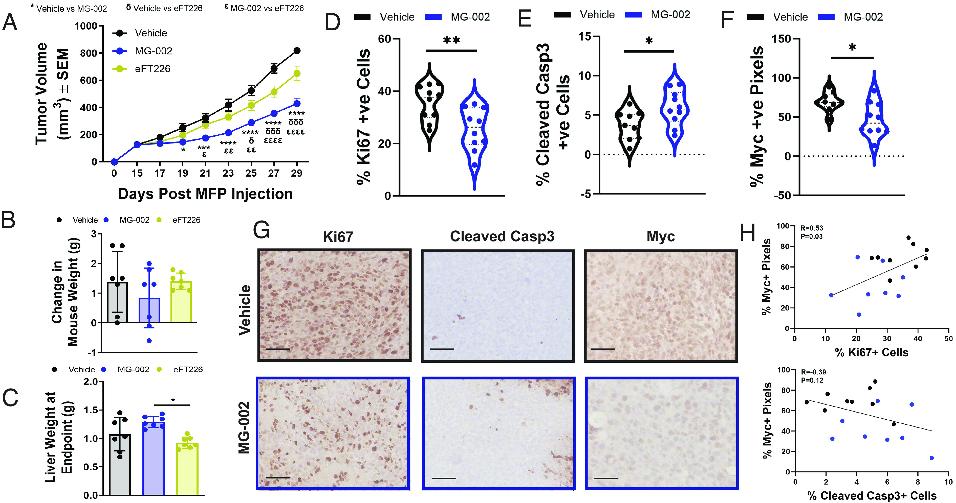
Fig. 5.

MG-002 inhibits primary TNBC tumor growth through reduced cell proliferation and increased apoptosis. (A) 4T1-526 mammary tumors were allowed to develop in the mammary fat pads of BALB/c mice and upon reaching ~100 mm3, the animals were randomized into three groups, which were treated by PO every 3 d with MG-002 (0.5 mg/kg), eFT226 (0.5 mg/kg) or vehicle control (n = 12 tumors/group). The data is shown as average tumor volume ± SEM. At the experimental endpoint, animals (n = 6 mice per group) were assessed for the (B) average change in mouse weight or (C) their liver weights. (D–F) IHC analysis of 4T1-526 tumors treated with 0.5 mg/kg MG-002 or vehicle control using (D) Ki67, (E) cleaved Caspase-3, and (F) Myc-specific antibodies. The data are shown as % positive cells ± SD and are representative of 9 to 10 tumors per group. (G) Representative IHC images for the graphs depicted in panel D–F. (scale bar: 100 microns.) (H) Pearson correlation examining the relationship between Myc positivity and either the % Ki67+ or % Cleaved Casp3+ cells (individual vehicle-treated tumors are represented by black dots; individual MG-002 treated tumors are represented by blue dots. For panel A, statistical analysis was performed with a two-way ANOVA (Tukey’s multiple comparisons test). For panels D–F, statistical analysis was performed using a two-tailed (unpaired) Student’s t test. *P < 0.05; **P < 0.01; ***P < 0.001; ****P < 0.0001.
