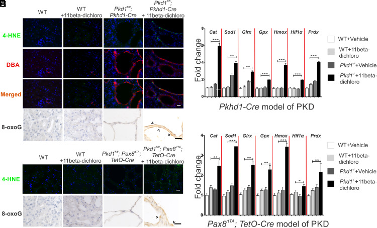
Fig. 4.
11Beta-dichloro exacerbates oxidative stress in cystic cells in both the early and adult mouse models of PKD. (A) In the Pkhd1-Cre model of PKD, the lipid peroxidation marker 4-HNE was used to examine the status of late ROS induction in WT and cystic epithelia in vehicle- vs. 11beta-dichloro-treated animals. The levels of 4-HNE (Top row) are specifically increased in the DBA-positive segments (second row) as compared with non-DBA stained tubules. Furthermore, WT mice treated with the drug from P10 to P23 did not display any overt 4-HNE signal (second column). (Scale bar, 20 µm.) (B) The oxidative stress biomarker 8-oxoguanine was probed in the same tissues as (A). Only 11beta-dichloro-treated cystic tissues displayed an increased signal. (C) For the early model, transcriptional levels of oxidative stress–inducible genes (Table 1) were examined via qPCR in wild-type and Pkd1−/− mice treated with vehicle or 11beta-dichloro. Total RNA from kidney tissue was collected at day P24; n = 5 for all groups. Only the cystic mice exposed to the drug displayed a specific activation of the indicated genes. (D) 4-HNE and (E) 8-oxoguanine staining in the adult Pax8 model of PKD. (Scale bar, 20 µm.) (F) For the adult Pax8 model, transcriptional levels of oxidative stress–inducible genes (Table 1) were examined via qPCR in wild-type and cystic mice treated with vehicle or 11beta-dichloro. Only the cystic mice exposed to the drug displayed a specific activation of the indicated genes. Total RNA from whole-kidney extracts was collected from mice at 5 mo of age; n = 5 for all groups. Statistics: N.S., not significant (P > 0.05); *P < 0.05; **P < 0.01; ***P < 0.001 determined by ANOVA. Data are plotted as mean ± SEM.

