Fig. 4.
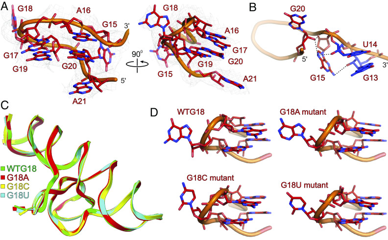
The structural features of the R domain and G18 mutant SCV PTE. (A) The overall structure of the SCV PTE’s R domain showing the flipped-out G18 (major binding site for the 5′ cap-binding protein, eIF4E). (B) Specific interactions of the G13, U14, G15, and G20 nucleotides within the R domain. (C) Superposition of the G18A (red), G18C (yellow), and G18U (cyan) mutant PTE crystal structures with the wild-type PTE. (D) Comparisons of the R domain structures for wild-type and G18 mutants showing that SCV PTE adopts a preorganized structure with flipped-out G18 that constitutes a ready-made platform for the eIF4E binding. Gray mesh represents the composite simulated anneal-omit 2|Fo|-|Fc| electron density map at contour level 1σ and carve radius 2.5 Å, and dotted black lines depict the heteroatoms within hydrogen bonding distances (≤3.0 Å).

