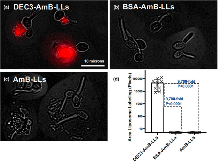
FIGURE 1.

DEC3‐AmB‐LLs bound efficiently to Candida albicans during the yeast–hyphal transition stage of development. (a–c) respectively, show representative photographic images of red fluorescent DEC3‐AmB‐LLs, BSA‐AmB‐LLs, and AmB‐LLs binding to C. albicans in the yeast–hyphal transition stage. After plating in media that stimulated hyphal development, yeast cells were grown for 1.5 h to reach this stage. The scale bar corresponds to 10 μm. Images were acquired at 60× magnification and equivalently cropped to enlarge cells for presentation. Composite images were prepared because the cells were widely dispersed in each 60× field. (d) The relative area of red fluorescent liposome binding (log10) was quantified using AreaPipe and is shown in scatter bar plots. N = 10 for each bar. Standard errors from the mean and p values are indicated.
