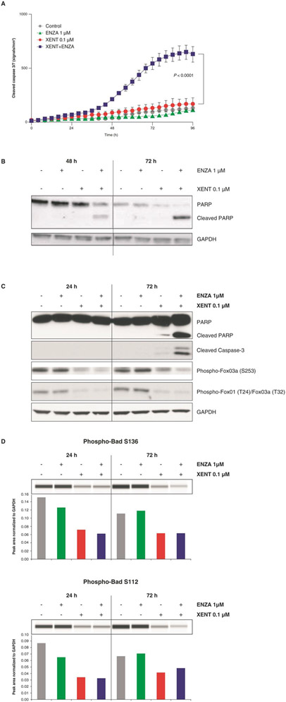
Figure 4.
Induction of apoptosis in VCaP cells: Effect of XENT and ENZA, alone or in combination, on caspase activity (A, C), cleaved PARP (B, C), phosphorylation status of FoxO3a/FoxO1 (C), and Bad (D). Cells were incubated with inhibitors as indicated in FBS-containing medium (without androgen or growth factor supplementation). Caspase-mediated apoptosis was detected using IncuCyte™ Caspase-3/7 Reagent and Western blot analysis. P values were calculated using pairwise t-tests (adjusted for multiplicity) following a one-way analysis of variance. Bad phosphorylation was determined by Simple Western™ quantification. All other proteins were assessed by Western blot analysis.

