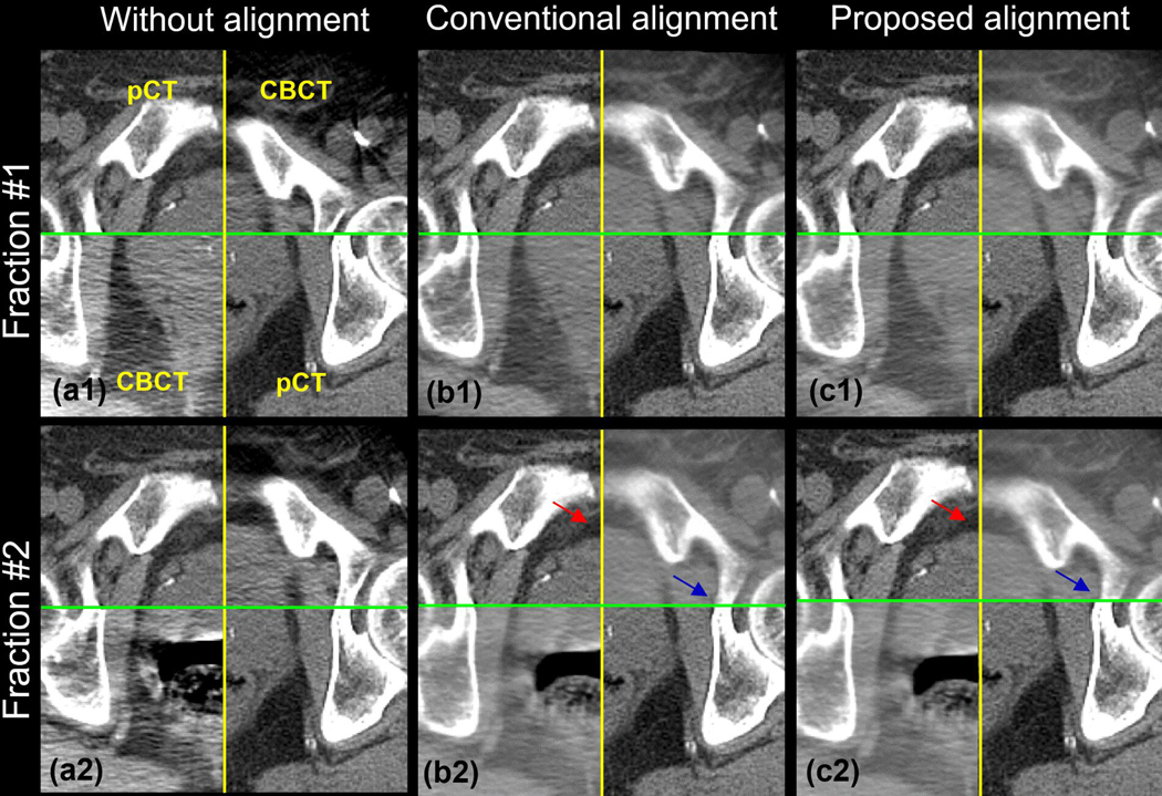
Figure 5.
The checkerboard of the FMs-free images without (a), with the conventional (b), and with the proposed (c) prostate alignment. Row 1 and 2 shows the result of the fraction #1 and #2, respectively. The relative motion between bony anatomy and prostate in fraction #1 is small, while fraction #2 is large.

