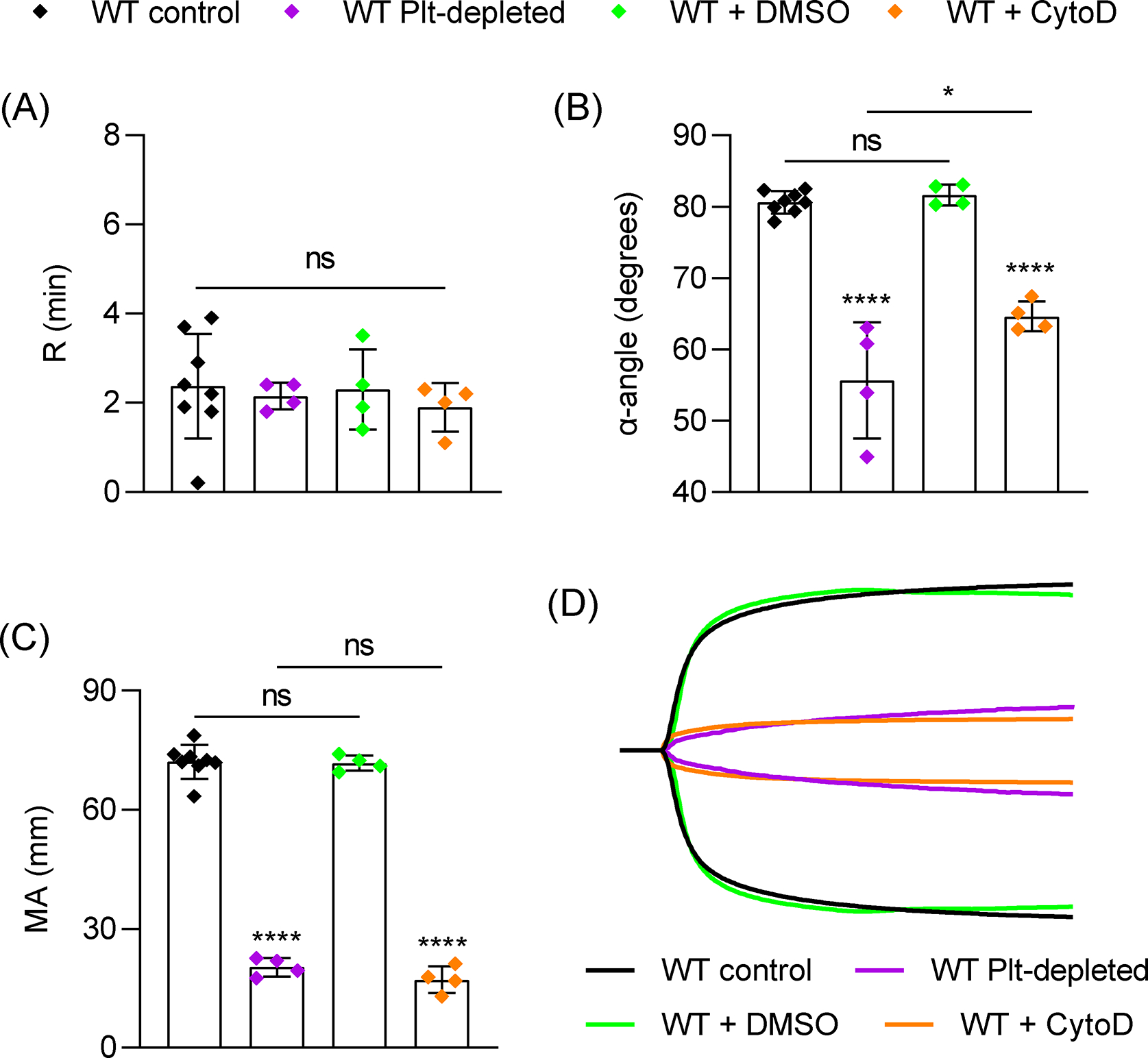
Figure 1:

Role of platelets and platelet contraction in TEG. Citrated blood samples were collected by retroorbital bleed (RO) from wild-type (WT) mice (n=8) or WT mice depleted of circulating platelets (WT Plt-depleted) by injection of an anti-GPIbα antibody (R300, 1 mg/kg, n=4). For inhibition of platelet-mediated contraction, WT samples were treated with DMSO (n=4) or cytochalasin D (5 μg/ml, n=4) for 10 mins prior to TEG assay. Samples were mixed with CaCl2 and kaolin in plastic TEG cup and run immediately in a TEG 5000 analyzer and recorded for 1 hour. (A-C) TEG parameters: R time (A), α-angle (B) and MA (C). (D) Representative TEG traces. Data shown as mean ± SD. Statistical significance was determined by unpaired Student’s t-test (A) or one-way ANOVA with Tukey’s multiple comparison test. Symbols directly over bars represent significance compared to WT control. *P < .05, ****P < .0001.
