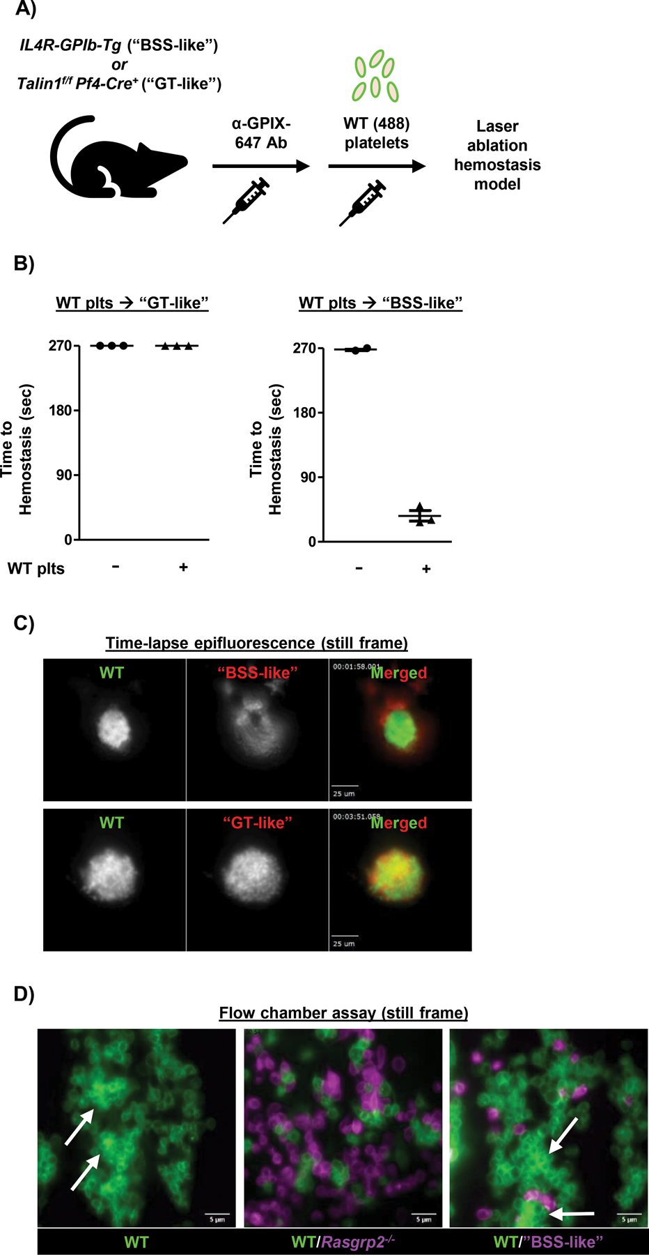
Fig. 4: Interference by dysfunctional platelets is dependent on GPIbα.

A) Model depicting platelet (plt) transfusion scheme. Endogenous plts in “GT-like” or “BSS-like” mice were labeled by injection of anti-GPIX-647 antibody prior to transfusion of GPIX-488 labeled WT plts. B) “GT-like” or “BSS-like” mice were transfused with WT plts to reach a circulating count of 1 × 10⁸/mL, for a ratio of ~ 7:1 endogenous:transfused plts in both mouse models. Time-to-hemostasis was then assessed in the laser injury model. n=3 per group. C) Mice were transfused with WT plts to reach a ratio ~ 2:1 endogenous:transfused plts, to allow for hemostatic plug formation in “GT-like” mice. Still frame images from epifluorescence videos (movies S9 and S11) demonstrate incorporation of “GT-like” (bottom panel) but not “BSS-like” (top panel) plts within the hemostatic plug. Some “BSS-like” plts can be seen adhered to the luminal side of the WT plug. Scale bars represent 25 μm. D) Flow chamber assay was performed on collagen-coated (200 μg/mL) coverslips at a shear rate of 1600 s-1. WT blood was flowed alone or after mixing at a 3:1 mutant:WT ratio with Rasgrp2−/− or “BSS-like” blood for 5 minutes over the collagen surface and visualized with a 100X oil objective. Plts were labeled with anti-GPIX antibody prior to mixing. Representative still frame images are shown. White arrows denote areas of WT thrombus formation. Scale bars represent 5 μm.
