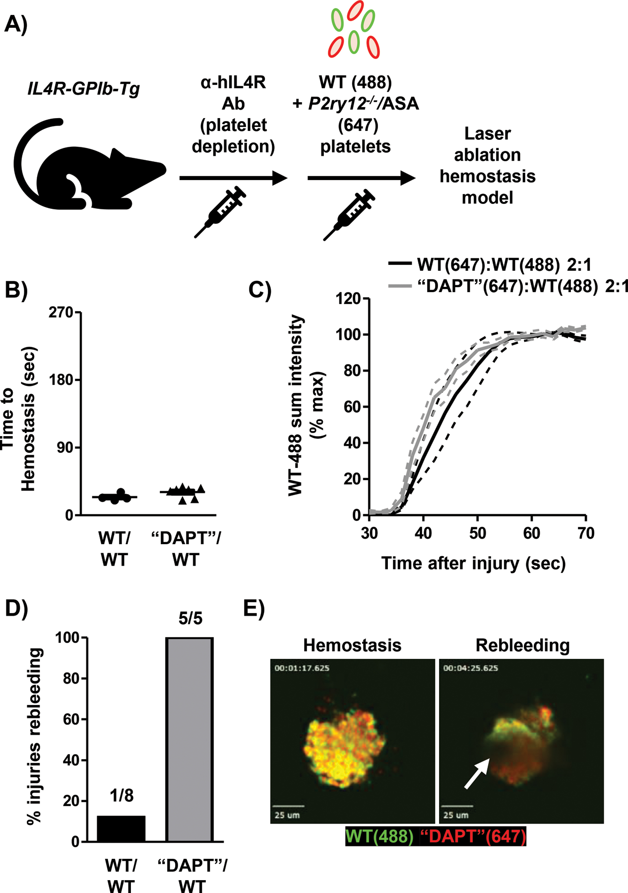
Fig. 6: “Dual anti-platelet therapy” platelets impair plug stability in the presence of WT platelets.

A) Model depicting platelet (plt) transfusion scheme. P2ry12−/− plts were treated with 2 mM acetylsalicylic acid (“DAPT”) ex vivo prior to transfusion. B) Time-to-hemostasis in TP Tg mice transfused with 2:1 “DAPT”:WT plts or 2:1 WT:WT plts was determined following laser injury. C) Adhesion of WT-488 plts was quantified in WT:WT or “DAPT”:WT plt transfused TP Tg mice. Data shown as mean ± SEM. D) Re-bleeding events (%) at individual injury sites during real-time SDC imaging. 5–8 injuries from 2–3 mice per group. E) Still frame images from real-time SDC video showing hemostasis and rebleeding at the same injury site (movie S13). White arrow indicates site of blood flow from hemostatic plug. Scale bars represent 25 μm.
