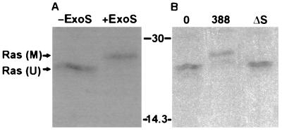
FIG. 1.
Mobility shift of Ras in HT29 cell lysates modified by ExoS in vitro and in vivo. (A) Ras modification in vitro by purified ExoS. HT29 cell monolayers were radiolabeled with [35S]methionine for 18 h and then lysed, and extracts were incubated for 30 min in the presence of buffer (−ExoS) or purified recombinant ExoS (+ExoS). Ras was then immunoprecipitated with monoclonal antibody Y13-259 coupled to anti-rat IgG plus protein A-Sepharose. Proteins were separated by SDS-PAGE and visualized by fluorography. M, modified Ras; U, unmodified Ras. (B) Ras modification in vivo by ExoS-producing bacteria. [35S]methionine-labeled HT29 cells were incubated for 3 h with McCoy’s-BSA alone (0) or with 108 CFU of ExoS-producing (388) or non-ExoS-producing (ΔS) bacterial strains as indicated. Bacteria were removed, cells were lysed, and Ras was immunoprecipitated and detected as described for panel A. Molecular masses (in kilodaltons) are indicated.

