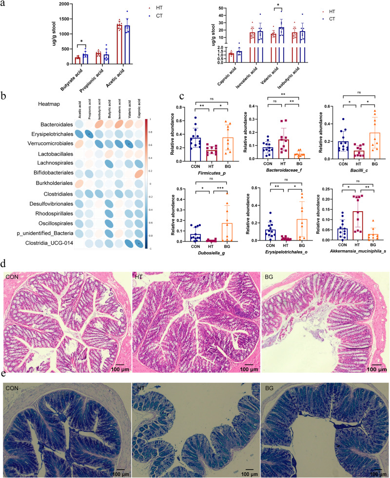
Fig. 5. Butyric acid altered the composition of gut microbiota of HT mice, and enhanced gut barrier integrity.
a The concentration of butyric, propionic, acetic, caproic, isovaleric, valeric, and isobutyric acids in fecal samples of the HT and control groups were determined by GC-MS. (N = 8 in each group). b Heatmap of correlations between the significantly changed gut microbiota and seven SCFA metabolites in HT mice at the family level. The color bar with numbers indicates the correlation coefficients. c Relative abundances of the following six significantly altered microbiota among the three groups: Firmicutes, Bacteroidaceae, Bacilli, Dubosiella, Erysipelotrichales, and Akkermansia_muciniphila. d Histological changes were observed using HE staining, and goblet cells are round cells that appear clear on HE staining and are typically flanked by the purplish absorptive-type cells. Scale bar: 100 μm. e AB/PAS staining on intestine tissue sections was performed to evaluate the histopathology of the intestines. N = 7 for CON group, 7 for HT group, and 8 for BG group. The Kruskal-Wallis test was used to detect significant changes for Bacilli, Dubosiella, Erysipelotrichales, and Akkermansia_muciniphila. The Welch’s ANOVA test was used for Firmicutes,and Bacteroidaceae. *P < 0.05, **P < 0.01, , and ***P < 0.001, ns, no significance.

