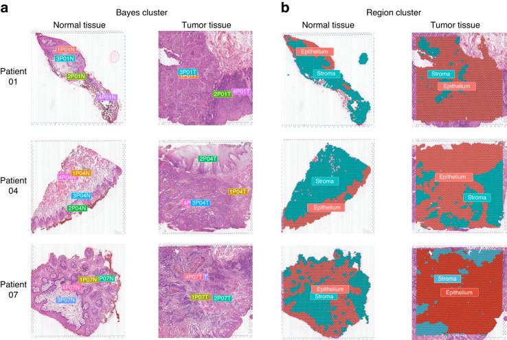
Fig. 1.
Spatial clustering of oral squamous cell carcinoma tissue samples. a Spatial clustering of BayesSpace was used to identify distinct regions within the tissue samples, and each sample was analyzed independently. b Based on the Bayes clusters, unsupervised clustering was used to divide the tissue regions into epithelial and stromal regions, which was confirmed by histological morphological information from hematoxylin & eosin (H&E) staining slides

