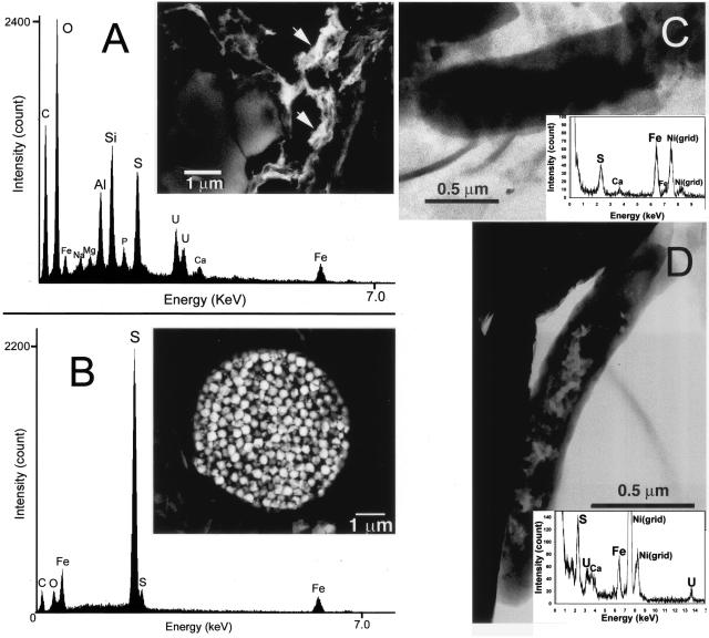
FIG. 2.
(A) Backscattered electron image of uranium-bearing phases (arrows) found in the aquatic sediment and an associated EDX spectrum. (B) Backscattered electron image of framboidal pyrite in the sediment and an associated EDX spectrum. (C) TEM image and EDX spectrum (∼30 nm spatial resolution) of a prokaryotic cell in the sediment that had accumulated iron and sulfur. (D) TEM image and EDX spectrum of a prokaryotic cell from the sediment that had accumulated uranium, iron, and sulfur.

