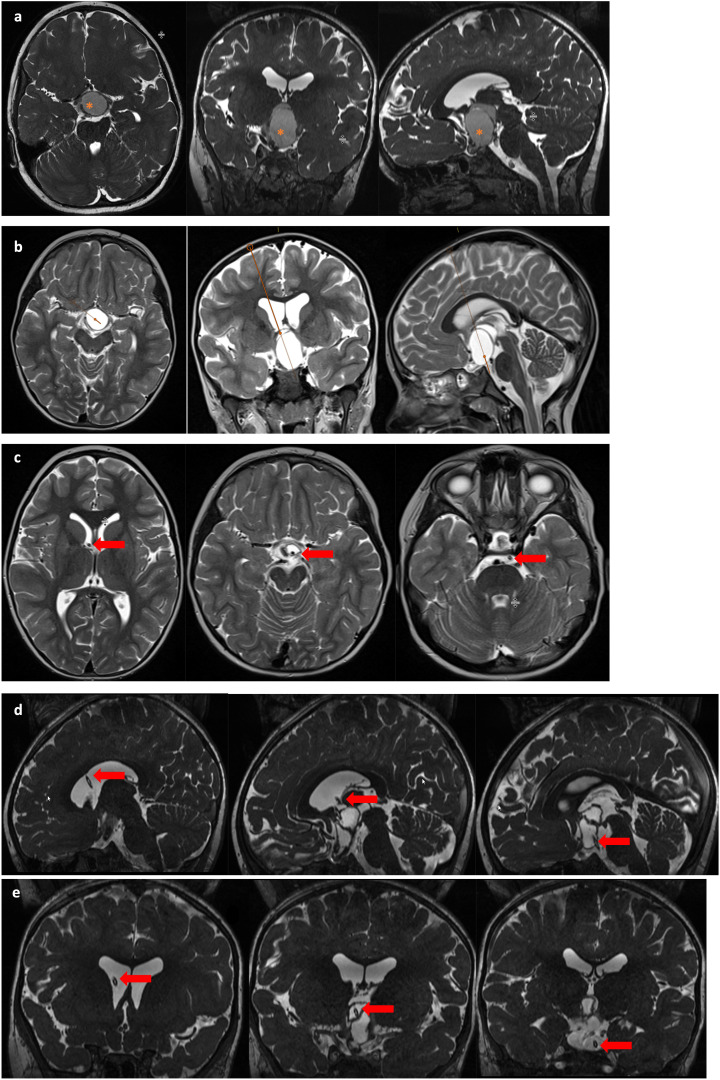
Figure 2.
MR imaging of a 5-year old female patient with a suprasellar cystic, space-occupying craniopharyngioma treated with stereotactic implantation of an internal shunt system. This patient presented with initial symptoms of mild visual deficiency and headache. Image line (A) demonstrates preoperative findings with a large intra-/suprasellar cystic tumor (*) compressing the chiasma. After planning the stereotactic trajectory (image line B) for implantation of an internal shunt system, the cystic tumor formation was reduced (image lines C (axial), D (sagittal) and coronal E) resulting in a decompressed chiasm and consecutive improvement of visual function. The arrow marks the course of the internal shunt through the foramen Monroi, the tumor cyst with final position in the prepontine cistern.

