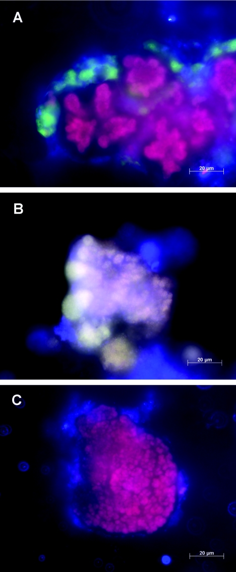
FIG. 2.
(A) FISH of an anammox enrichment sample from a 2L laboratory reactor containing mostly “Ca. Kuenenia stuttgartiensis” with probe S-*-Kst-1275-a-A-20 (labeled with Cy3; red) and probe S-*-Amx-0820-a-A-22 (labeled with Cy5; blue). Overlapping red and blue labels result in purple “Ca. Kuenenia stuttgartiensis” cells. Remaining blue cells might be another type of anammox bacteria. Autofluorescence is depicted in green. (B) FISH of an anammox biomass sample containing “Ca. Scalindua” with probe S-*-Amx-0368-a-A-18 (labeled with Cy3; red), probe S-*-BS-820-a-A-22 (labeled withFluos; green), and the Eub probe mix (7) (labeled with Cy5; blue). Overlapping red, green, and blue labels result in anammox organisms that appear yellow-white. (C) ISR-FISH of an anammox enrichment sample from a 2-liter laboratory reactor containing “Ca. Brocadia anammoxidans” with the probe mix targeting the ISR of “Ca. Brocadia anammoxidans” (labeled with Cy3; red) and probe S-*-Amx-0820-a-A-22 (labeled with Cy5; blue). Overlapping red and blue labels result in anammox organisms that appear purple.

