Fig. 5.
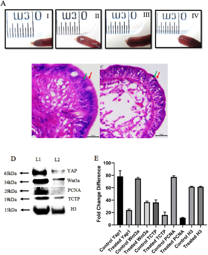
TCTP and its connecting link with regenerative protein: A (I). Control amputated worm with 5th day bud. II, III and IV represent progressive regeneration suppression in Nutlin-3a injected worms with 5, 7, and 9 µg, respectively. B Histology of control 5th -day regenerative blastema with well-organized tissue structures has the most thickened outer and inner epithelial layer. C In Nutlin-3a injected worms, the 5th day bud is not well organized, loosely packed with internal bud tissues and thinner layers of the outer and inner epithelium. D Western blotting image represents that when compared to the control samples (7th day regeneration—Lane 1), all frame of regenerative key proteins was notably reduced in Nutlin-3a treated samples (7th day regeneration—Lane 2). Nutlin-3a is known for TCTP silencing, and according to the result, TCTP silence influences organ formation (YAP1), stem cell activation (Wnt3a) and cell proliferation (PCNA). E. Quantification of YAP1, Wnt3a, TCTP, PCNA and H3 expression are done based on the band intensity and represented using bar diagram. The experiments were repeated in triplicate to analyze the statistical significance, representing their value as mean ± SD. p value < 0.05 was considered statistically significant data. The red arrow represents the “Outermost epithelial layer”; the Green arrow represents the “Inner layer of epithelial tissue.”

