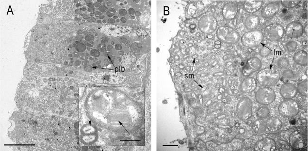
FIG. 1.
Transmission electron micrographs of Bathymodiolus sp. gill sections with endosymbiotic bacteria. (A) Transverse section showing an overview of a bacteriocyte (plb, phagolysosome-like bodies). Scale bar = 10 μm. (Inset) Large morphotype with stacked internal membranes (arrow) and a dividing stage of the small morphotype (arrowhead). Scale bar = 0.5 μm. (B) Apical part of a bacteriocyte showing the distinct distribution of the two morphotypes. The smaller morphotype (sm) occupies the apical part of the bacteriocyte toward the mantle fluid, while the larger morphotype (lm) is located more basally. Scale bar = 1 μm.

