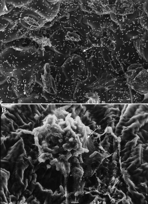
FIG. 1.
Scanning electron microscopic micrographs of field-grown bean leaf surfaces colonized by seed-borne X. axonopodis pv. phaseoli. (A) Leaf surface showing mostly solitary bacterial populations (Sol. b.). Note the accumulation of bacterial cells in grooves (G) between epidermal cells. Bar, 10 μm. (B) Focus on a bacterial biofilm. Note the matrix (M) embedding bacterial cells constituting a typical biofilm. Bar, 1 μm.

