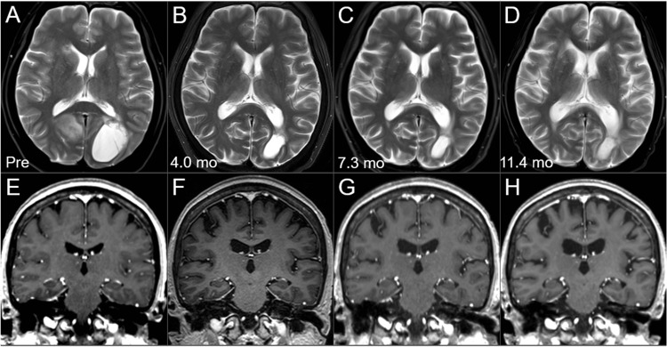
Figure 9. Morphological changes of the brain on magnetic resonance images before and after the external beam radiotherapy for brain metastases.
The images show axial T2-WIs (A-D) and coronal CE T1-WIs (E-H); before the WBRT (pre, at age 57) (A, E); at 4 months after the WBRT (B, F); at 7.3 months (C, G); and at 11.4 months (D, H).
(A-H) These images are shown at the same magnification and coordinates under co-registration and fusions based on the images before the WBRT. In particular, when comparing the images before and 11.4 months after the irradiation, ventricular dilatations, slightly high-intensity change in the periventricular deep white matters, widening of the cortical sulci, and the Sylvian fissures, suggesting the progression of parenchymal atrophy and degeneration, are observed, which are difficult to explain by tumor shrinkage alone.
WI: weighted images; CE: contrast-enhanced; WBRT: whole-brain radiotherapy

